CN
10x Xenium

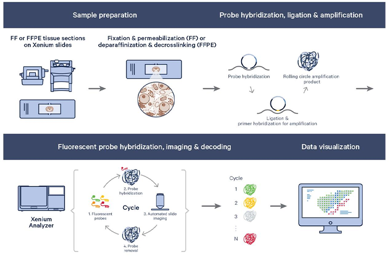
Xenium technology adsorbs a DNA probe to the location of the target mRNA on the slice by hybridizing an in situ nucleic acid probe to the slice, coupled with an mRNA-dependent DNA ligation reaction. Hybridizable sites are increased by rolling loop replication of the probes. The samples are then hybridized by multiple rounds of multicolor fluorescently labeled oligonucleotides, and the spatial fluorescence signals of each round are recorded with a microfluorescence scanner. The spatial distribution of the target mRNA is detected by the color information of the spatial fluorescence signals in multiple rounds.

Service Advantage

Service Advantage

High sensitivity and specificity

The unique padlock probe design enhances the sensitivity and specificity of the assay.

High resolution

Combined with the detection method of in situ microimaging, in situ gene expression analysis at the subcellular level is realized, which greatly improves the spatial histological resolution.

High throughput

The Xenium slide has an imaging area of 12mm x 24mm, allowing two slides to be examined in parallel at one time; while meeting the needs of tissue studies with large cross-sections, it can also be used to examine multiple tissue sections on a single slide; at present, hundreds of target genes can be analyzed on each slide, and 1,000 target genes can be examined at a later stage.

High flexibility

A variety of pre-built panels are available, and up to 100 customized target genes can be added to meet the individual needs of a specific study.

Non-destructive

Xenium's non-destructive analysis maintains histomorphometric integrity post-run, allowing RNA, IF, and morphology (H&E) information to be obtained on the same fresh frozen or FFPE section for seamless integration with histopathology studies.

Sample Types

Panel

Customised
Services

COPYRIGHT©Infinity Scope Multi-Omics Biotechnology Co. Ltd., All rights reserved. 浙ICP备2023015471号-1