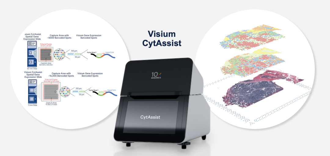
In the Visium CytAssist workflow, sectioning, tissue preparation, staining (H&E or IF) and imaging are all performed on standard slides. After probe hybridization, two standard slides and one Visium slide with two capture areas are placed into the CytAssist instrument, aligning the tissue sections on the standard slides with the capture areas. The instrument captures a bright-field image to provide spatial localization for data analysis, and the transcriptome probe is subsequently hybridized to the Visium slide.

1. paraformaldehyde (PFA)-fixed frozen tissue, fresh frozen tissue, or formalin-fixed paraffin-embedded (FFPE) tissue
2. Human or mouse samples
3. different sizes of tissue sections, flexible analysis of up to 11 x 11 mm region of interest
COPYRIGHT©Infinity Scope Multi-Omics Biotechnology Co. Ltd., All rights reserved. 浙ICP备2024079019号-1